Introduction
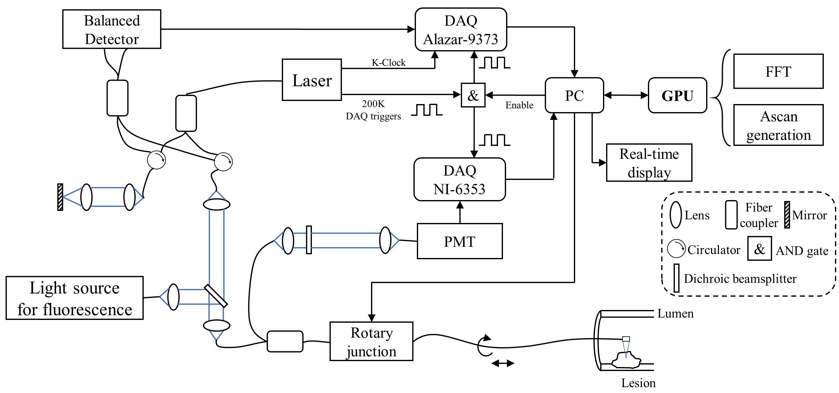
Near-infrared light has limited penetration into human tissue. To image internal organs, we require a miniature probe to help the imaging system reach deeper into the body. By using a catheter and a rotary motor, the OCT system can image internal tissue structures, such as those in the cardiovascular. Clinically, in addition to structural imaging, it's also crucial to observe biological processes. Fluorescence imaging leverages either the autofluorescence of biological tissues or the fluorescence of dyes to perform molecular imaging, thus enabling observation of living organisms' life activities. This project achieves in vivo multimodal imaging by combining OCT with fluorescence imaging, allowing us to capture both structural and molecular information. Additionally, with the aid of an endoscopic catheter, we can image the internal tissues of humans and animals, such as the colon of a mouse.