Introduction

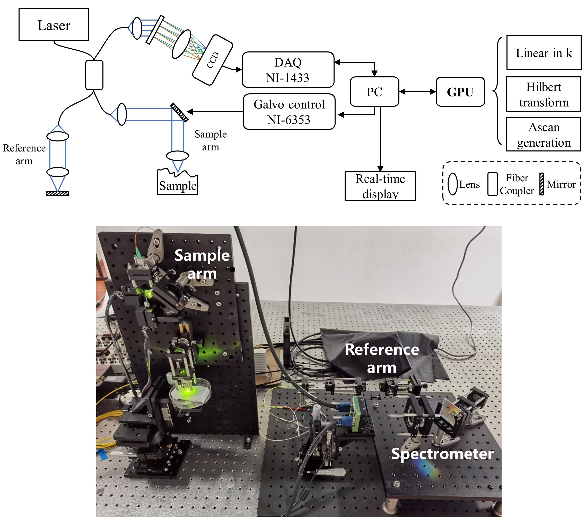
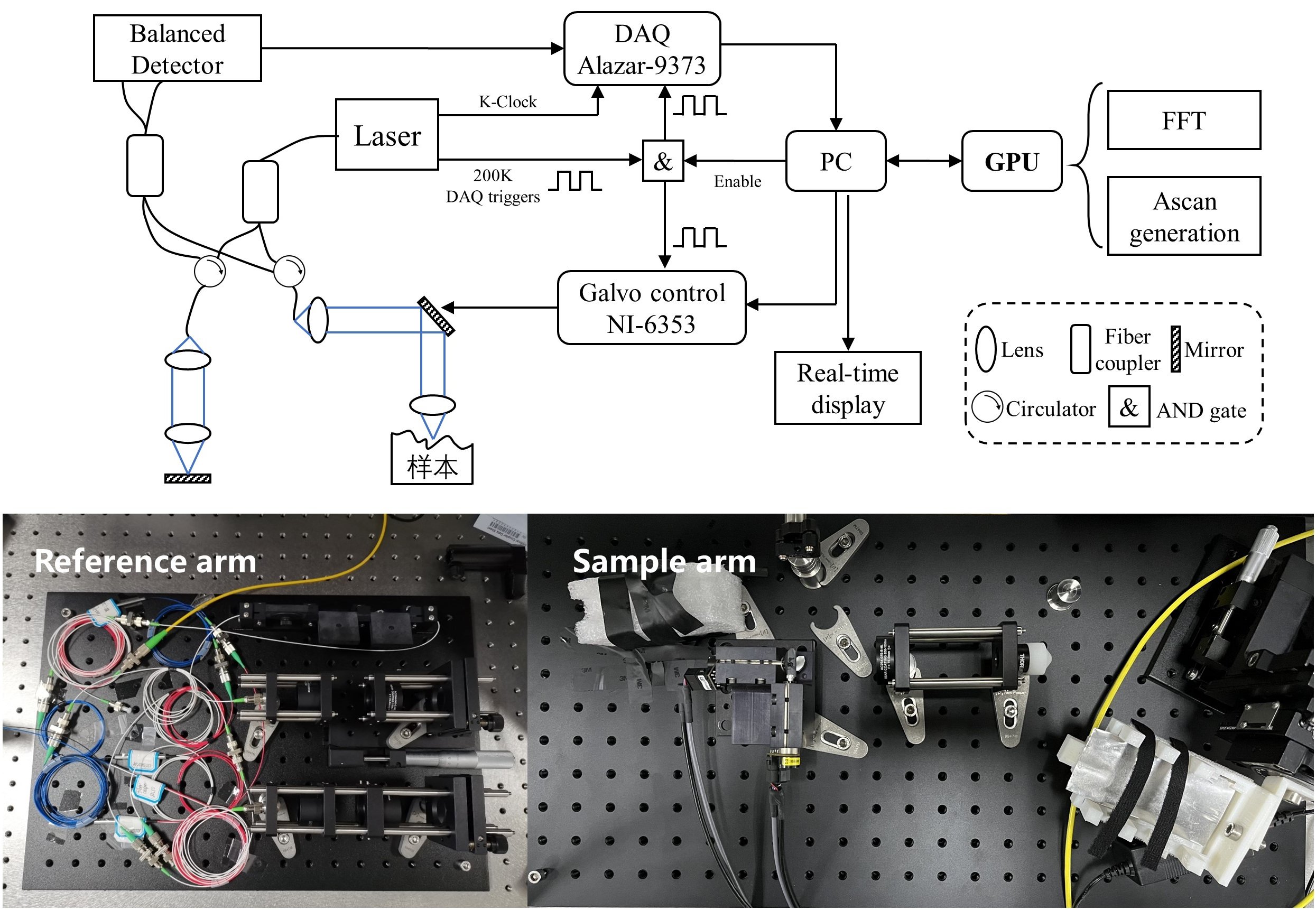
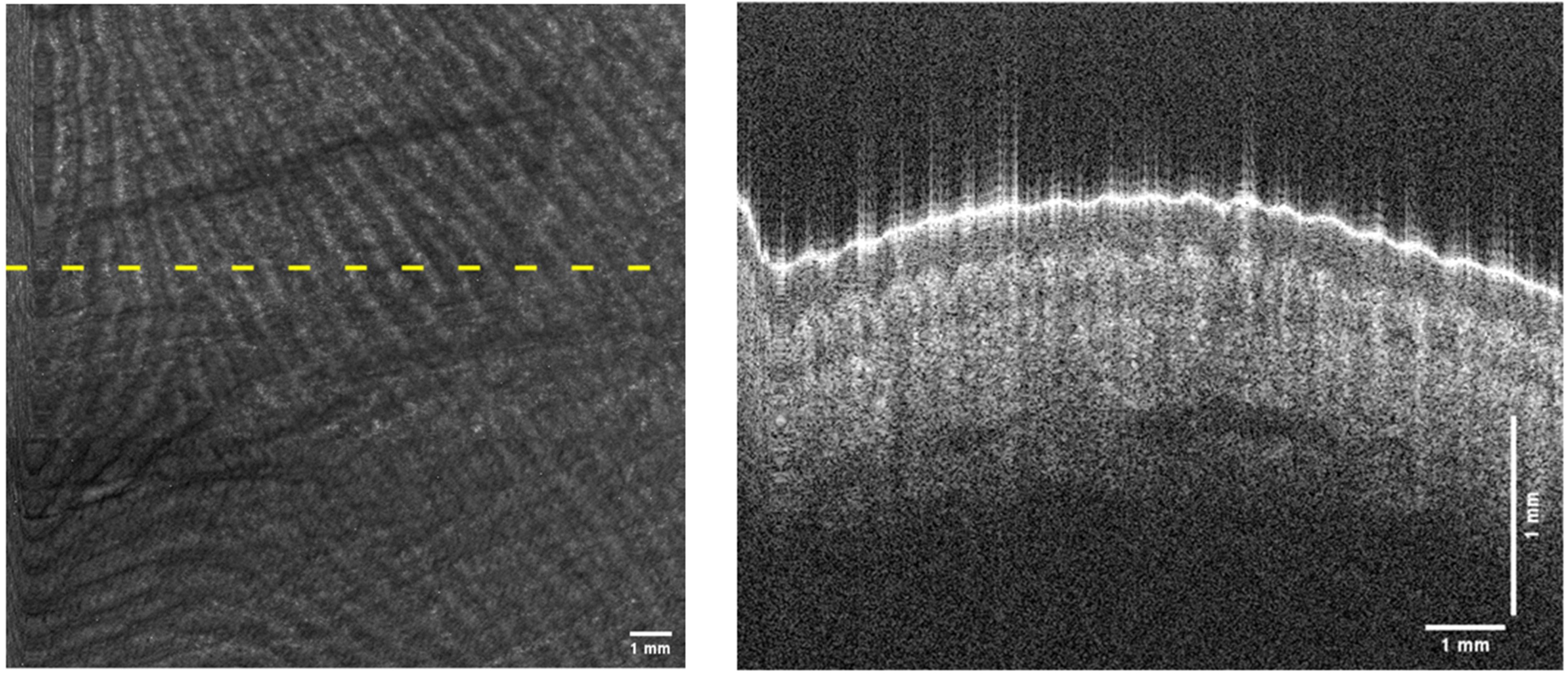
Optical Coherence Tomography (OCT) is a 3D imaging technique with millimeter-level imaging depth, micrometer-level resolution, and KHz to MHz imaging speed. It has become the gold standard in ophthalmology and has been used hundreds of millions of times in clinics worldwide. In this study, we built near-infrared swept-source OCT (SSOCT) and spectral-domain OCT (SDOCT) systems, and successfully imaged human fingerprints, human fundus, mouse colon, mouse retina, porcine coronary artery, and other biological tissues.
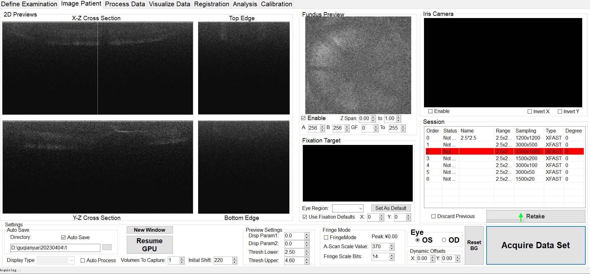
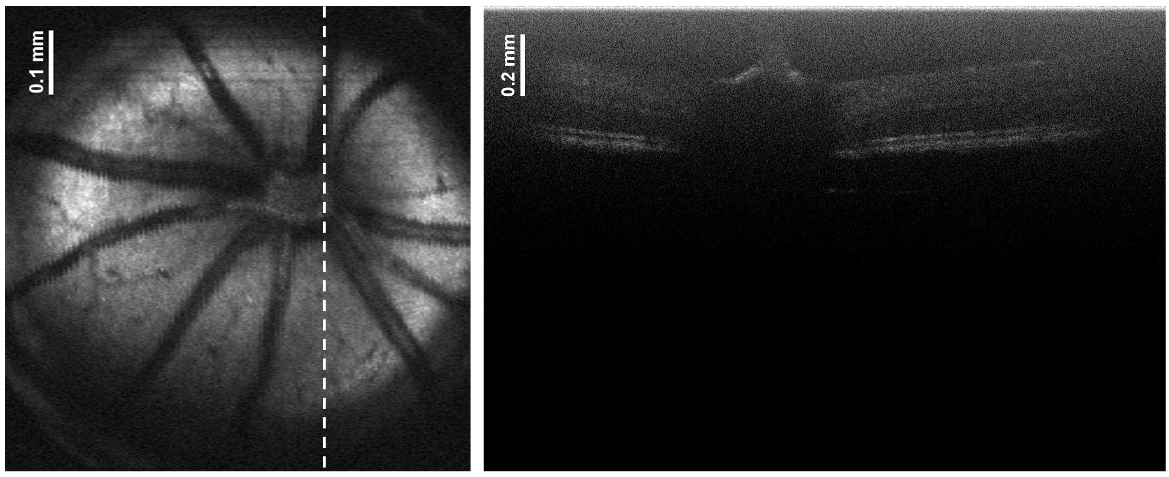
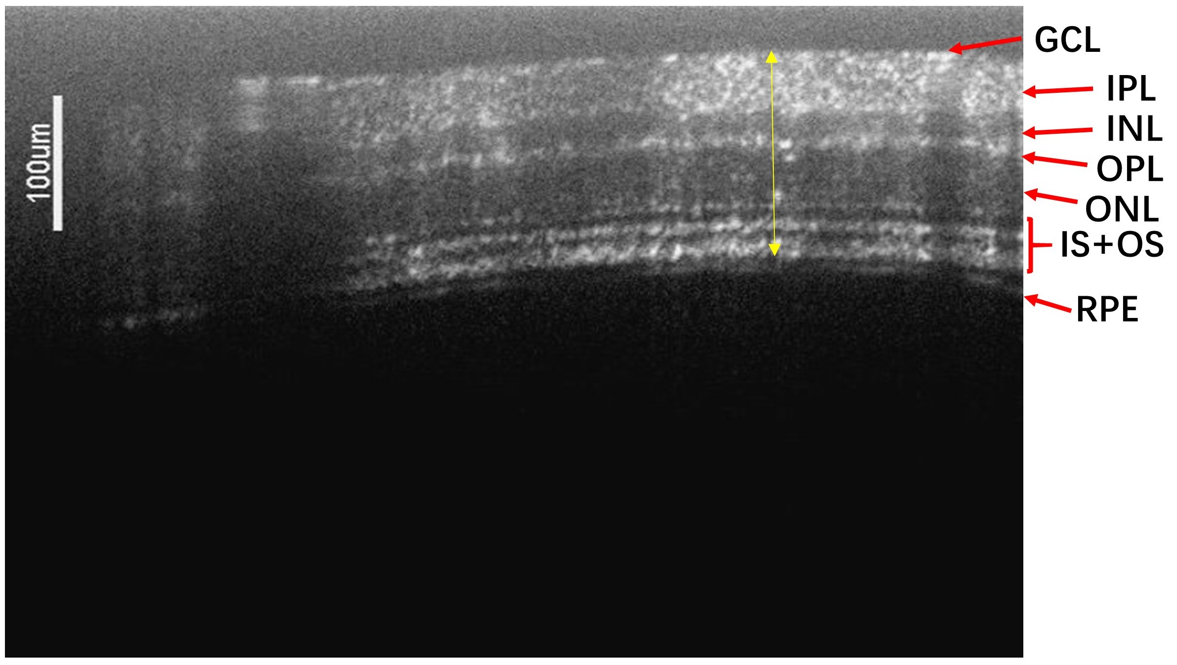
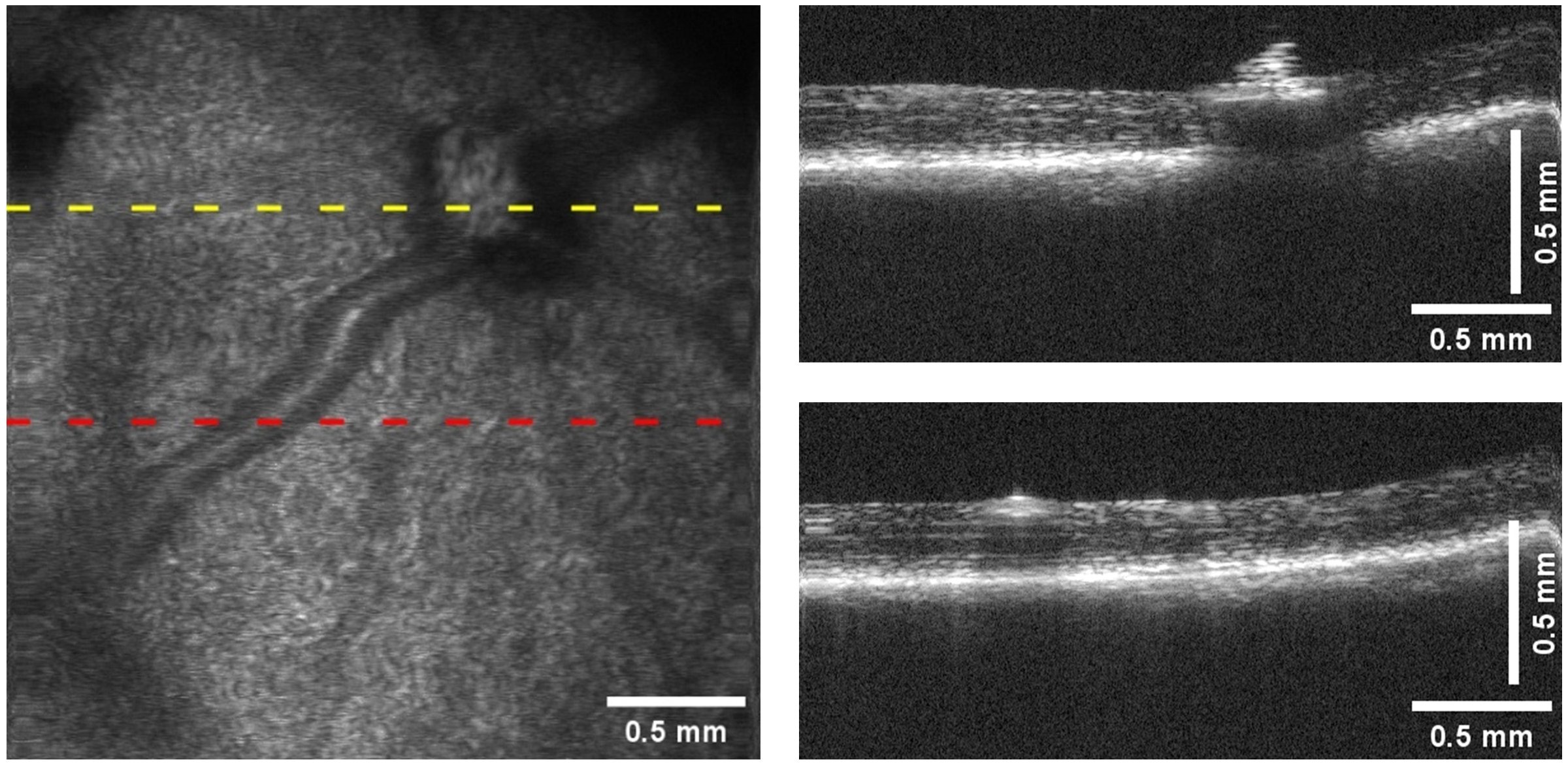
Retinal diseases are serious conditions that significantly impact people's quality of life. The prevention and treatment of these diseases pose an international challenge, largely due to unknown pathogenic mechanisms. Current research relies heavily on mouse models, and OCT technology plays a crucial role in observing their retinal structures. However, the rapid onset of cataracts in experiments complicates the identification of the optic nerve head. In this study, the use of GPU acceleration technology gives both OCT systems real-time imaging capabilities, enabling us to locate the optic nerve head more efficiently. This advancement addresses a significant need in retinal disease research.